Sunday, August 8, 2010

51. When to operate on a closed pyometra case?

SUNDAY'S CASE

August 8,2010

Sunday, August 8, 2010

Consultation at Vet 1.
The 9-year-old, 5-kg Jack Russell passed out some sticky vaginal discharge and also blood in the stools on Saturday. The young lady and her brother googled "pet clinic" and went to North-east Singapore.

Vet 1 shaved bald the dog's ventral area for an abdominal ultrasound scan. "Lumps of around 3 cm in diameter can be seen", he told the owner. Vet 1 diagnosed pyometra based on vaginal discharge, fever, abdominal palpation and scan. Surgery and anaesthesia quoted was $1,000.

The kind vet advised surgery as soon as possible and presented the young lady a list of vet practices in Singapore. She was living in Toa Payoh and phoned me for a quotation. I was surprised when she told me that she did not know of Toa Payoh Vets existed (since 1982) although she is a Toa Payoh resident.

CHANCES OF SURVIVAL
It is difficult to guarantee 100% survival. The dog had actually been vomiting occasionally in the past 3 months, not just last week.

Vet 1 gave a Rimadyl 0.5 ml (50mg/ml) injection 2 hours ago. The temperature before Rimadyl injection was 39.7 deg C (fever). When I checked the dog 2 hours later, it was 38.5 deg C (normal). So I had to decide to operated soon as the fever would return when the drug effect went off.

I gave an antibiotic (Baytril) 0.6 ml SC 1 hour before surgery. The dog was operated and recovered well.


She was still drowsy when the young lady and her brother visited her 2 hours after surgery but could recognise them.
I showed the pyometra to the young lady and her brother. "Vet 1 said it was 3-cm lumps," she said. "The ultra-sound cannot measure all lumps accurately," I said. "Some of the lumps in this uterine body are 3 cm in diameter and some are more or less." I told her. She was very relieved that her dog was alive and that was what counted.

RIMADYL (a class of pain-killer called NSAID).

1. I have never used Rimadyl before surgery although the manufacturer (Pfizer's website) does recommend it, at 2 hours before surgery so that the dog will not feel any pain after surgery.

2. The manufacturer advised the use of parenteral fluids during surgery to reduce the potential risk of renal complications when NSAID is use perioperatively.

3. In this case, the first vet had given 0.5 ml Rimadyl (50 mg/ml) SC 2 hours ago and no antibiotics. The dog had a fever of 39.7C. When I was consulted 2 hours later, I checked the temperature. It was 38.5C. The owner commented that the dog looked better. Rimadyl had reduced the pain. It also appeared to be anti-fever too.

Calculation of dosage in this case.
Jack Russell, Female, 9 years old, 5 kg.
Manufacturer advised: 4.4mg/kg for 24 hours or 2.2mg/kg two times a day by injection. Same dosage for oral administration.
Therefore, the first vet gave 25 mg SC or 5 mg/kg.

I decided to operate on the dog 2 hours after consultation as the fever had subsided and might return if there was a delay. The dog growled after some time when I checked her gums which were pink but dry-looking. I could feel a large swelling in her lower 1/3 of the abdomen but not the lobes felt by the first vet. She looked "pregnant". Rimadyl might have made abdominal palpation difficult. There was no pain reaction from this Jack Russell.

Rimadyl provides pain relief and was already given by the first vet during consultation. I did not give anymore for the next 24 hours.

SEVERE DRUG REACTIONS ASSOCIATED WITH NSAIDs
include gastrointestinal, renal and hepatic signs and in rare situations, death.

Therefore, in pyometra post-surgery, I don't give NSAID before or after surgery as I don't know how badly damaged the dog's kidneys are. This dog had been vomiting for the last 3 months and would not be considered healthy.

It is safer as the owner wants a dog alive at the end of the surgery.


photos and updates are at www.toapayohvets.com

Friday, August 6, 2010

50. Right perineal hernia repair in the Boston Terrier

Today is Saturday, August 7, 2010. It is my day off but I better record my recent surgery on the right side of the backside of the Boston Terrier before I forget as there are so many things to do and read in Singapore as compared to Perth, Australia where the pace of life is much less hectic.

"The backside lump keeps getting bigger," the young man and caregiver of the Boston Terrier phoned me in the first week of August 2010. "My dog can't poop normally. You need to operate on him."

I had repaired the left perineal hernia in a Boston Terrier in Jun 2010. The surgical outcome was excellent and I was glad. Surgeries take a long time and can result in complications.


Left perineal hernia in a Boston Terrier has been repaired in June 2010
The right perineal hernia started to enlarge bigger and bigger

"The swelling is the rectum full of stools. Part of the rectum had dropped into this perineal hernia and you can see it as a lump. It is therefore hard for the dog to pass motion. Use your finger to push the lump back into the abdomen so that he can poop and make an appointment for surgery," I had just neutered this dog 7 days ago. Neutering is reported to reduce the incidence of perineal hernias in the male dog. It was better late than never.

ANAESTHESIA
The owner is worried that his old dog may not survive a 3rd anaesthesia. The first two were for the repair of the left perineal hernia in June 2010 and neutering. Many dog owners in Singapore are aware of the risk of the dog dying on the operating table and the internet forums of such cases frighten them. In this dog, the only cure was to operate to close the big hole inside the right perineum as the dog had great difficulty and pain in pooping.

Domitor 0.2 ml IV and isoflurane gas. An I/V saline drip. No problem. I did not use Antisedan to wake up the dog as he would wake up naturally.

SURGERY
The hole was even larger than the left perineal hernia I did in June 2010. My assistant Mr Saw wore a glove and asked me: "Do you want me to dig out the stools?" I said: "It is better not to do it. The dog's rectum may be traumatised. In any case, it is not necessary to empty the stools from the rectum." I was never more mistaken.

Under anaesthesia, the solid stools started to pop out of the anus. My assistant removed them with a tissue paper. I had not plugged the anal opening with the syringe or something.

I used electro-incision to open a big curved incision of the skin. Fatty mass spilled out of the wound. I used the retractor to open widely and horizontally the herniated muscles.

I could not believe it. The whole defect was over 4 cm x 6 cm in diameter and very deep. As deep as 3 cm. There was not much muscle layer to stitch from the right lateral side to the anal sphincter. Would the stitches hold? There would be no second chances.


Right perineal hernia in a Boston Terrier has been repaired in August 2010
In this case, I could see the reddish right ventral wall of the rectum. I placed several layers of 2/0 absorbable stitches to close the gap. "The retractor is very useful," Mr Saw remarked. "Yes," I said. "It opens up the hole so that I can stitch up properly."

The whole area was a mess. I could see one big vein but not the nerve which was supposed to be present in theory.

POST-OP CARE
In theory, the dog should be crated for 14 days and given limited movements for another 14 days so that the stitches would not break down. To save money, the owner would take the dog home in 4 days and confine the dog himself.

CONCLUSION
Sometimes it is better to neuter the dog when he is young. For the past years, I have had not seen a case of perineal hernia in neutered dogs. The incidence in neutered and female dogs is very low. In just the past 2 months, I have had operated on 2 male not neutered dogs with perineal hernias.

One of the recent cases of a right perineal hernia in an 8-year-old Shih Tzu is at:
http://www.asiahomes.com/singaporetpvet/0514haematoma_perineal.htm


Right perineal hernia in an 8-year-old non-neutered Shih Tzu. The hernia was discovered by the teenaged girl "recently". The complaint was "constipation"
Perineal hernias are not common conditions in the sense that I don't see them every week. But they are commonly present in non-neutered dogs. When you see a swelling to the right or left of the anus, get your vet to quickly repair the perineal hernia rather than wait for a long time as it is extremely difficult to stitch up a big gap in the pelvic diaphragm compared to a small one.


Pictures are at www.toapayohvets.com, goto DOGS

Thursday, June 24, 2010

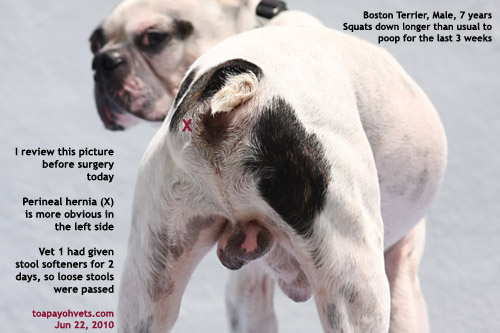
49. Perineal hernia in a male Boston Terrier

A knowledgeable young man who has his own views of dog care.
He presented a Boston Terrier with constipation for the past 3 weeks. He went to Vet 1 who referred him to another vet after taking a blood test as she did not want to perform the surgery. She had given him a laxative for the dog and the dog had passed loose stools instead of hard ones.

"Why don't you see the referred vet?" I asked.
"The Surgery had a bad reputation," he had googled the name of the practice and there was a nasty complaint from one dog owner.
"All practices will have one or two bad complaints about service," I said. "The busier the practice, the higher the chances of getting complaints as the vet may have no time to handle each case. This applies to over-worked doctors in human medicine too especially in the emergency wards."

"Has your dog been vaccinated?" I asked.
"No vaccination for the past few years. Do you have parvovirus in your Surgery?" the young man asked me.
"Fortunately for your dog, my practice does not have parvo-viral cases for many months as I seldom provide service to the dog breeders nowadays. You have taken a big risk exposing your dog the risk of parvoviral and canine distemper infections."

PRE-OP BLOOD TEST IMPORTANT FOR OLD DOGS BEFORE SURGERY
I checked Vet 1's blood test results. It is wise not to trust the blood results of other practices based on one of my experiences (see one case I had written). However, he had paid $130 for the test and I would not insist as that would increase his vet bills. Overall, the dog was examined and was healthy. So I took the chance.


ANAESTHESIA
Domitor and isoflurane gas was OK. The dog recovered smoothly.

SURGERY
I gave him antibiotics to take and schedule perineal hernia surgery 2 days later. The surgery took nearly an hour as the hernia was large.


CONCLUSION
The dog was OK and was warded for at least 4 days as the owner did not have a crate to prevent the dog running loose. I checked the dog every day to ensure that he had proper nursing care and pain-killers. The boy's parents came to visit the dog yesterday. The dog should be back home after 7 days. He had managed to rub his backside onto the floor of the crate despite tolfedine 60 mg at half a tablet per day for 3 days. I decided to give him 1/4 dose of a 30mg phenobarb and then rimadyrl for another 3 days to prevent pain and inflammation.

Updates at www.toapayohvets.com

Tuesday, March 2, 2010

48. Serious heart disease + deep eye ulcer


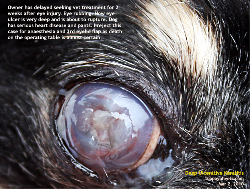
The mother and teenaged daughter adopted the abandoned Chihuahua. For the past 2 weeks, the dog was rubbing her eyes. Dirt and bacteria get trapped inside the eyes, irritating the dog. So, she rubbed more. When I was consulted, the eye cornea was deeply ulcerated. A 3rd eyelid flap would be the surgery and would have saved his eye sight if the owner had not delayed seeking vet treatment.

In addition, this Chihuahua had a serious heart disease. Panting when at rest. More panting when excited. A pre-anaesthetic sedation using Domitor might be too risky.

Therefore, I did not proceed with the surgery as she would likely just die on the operating table. A 90% chance of dying. The owner accepted the risk but since this is a very high risk, I did not want to risk my reputation in handling this case.

The dog went home with heart drug medication, antibiotics, eye ointment and an e-collar. It was better to have a live pet as the teenaged daughter was attached to her. No vet wants a dead dog as a result of anaesthesia. No owner wants that too.

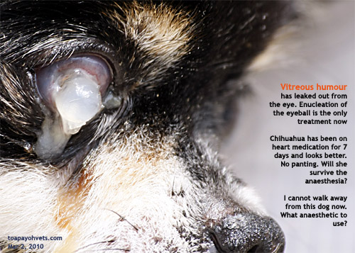
One week later, the eye ulcer had perforated. A white plug of gel oozed out. What to do now? Bacteria would get inside the eye causing enopthalmitis. Uveitis was controlled by eye drops. There was one solution. Surgery to remove the whole eyeball (enucleation).

Performance counts. In this case, a good performance is a live dog at the end of the surgery. The following management of this case is shared with readers and vets:

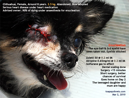
1. Dog was already on Fortekor for the past 7 days. She was no longer panting but dislike direct mask isoflurane gas. Struggled. I did not risk masking her since she might just collapse of heart failure during the fight to get out of the mask. So, a sedation injection was needed. Domitor or zoletil? Which is safer? Does Domitor cause heart rate to slow down? How about xylazine? It causes hypotension. Eventually, I used Zoletil. But how little that can be effective is needed for this 2.5 kg dog?

2. Pre-anaesthetic dextrose saline 20 ml SC and antibiotics.

3. Weight was 2.5 kg. Thin.
Pre-anaesthetic dose of Zoletil 50 @ 0.2 ml IM.
Atropine 0.65mg/ml @ 0.2 ml IM to prevent salivation, neck twisting from Zoletil injection.
Isoflurane gas mask and then intubate.
4. The dog's eyelids were shaved fast. The eyeball was taken out in <10 minutes.

5. The teeth were scaled. 4 loose ones extracted.

6. The dog was on <1% isoflurane gas and was anaesthesized without problems. For a short period, I disconnected the gas and the dog was not awake. So, I could continue surgery. When the dog moved, I re-connect the gas. This close observation and not giving too much anaesthesia ensures the survival of the patient.


7. Post-op dextrose saline 20 ml SC, antibiotics and tolfedine painkillers.

8. Dog went home on Day 2. No problems. Everybody is happy when a dog is alive at the end of surgery and anaesthesia. If the dog had been operated earlier, she would have had died on the operating table as she never had been checked up for heart disease and was not stablised on heart medication. Pictures of the eye injuries are below.

Pic at www.toapayohvets.com

Saturday, February 13, 2010

47. Oxytocin v. elective Caesarean section

If the female dog can't give birth naturally, give an injection of oxytocin and see what happens. This is fine in theory and in lectures.

In private practice, the owner wants a good outcome when he brings in a barrel-shaped Miniature Schnauzer that had difficulty in giving birth to the vet for Caesarean section. With a fully distended abdomen, the Schnauzer is likely to have 5 or 6 puppies. In this case, an X-ray by a vet said 4 puppies but the owner did not bring the X-ray.

Now, what the owner wants is that he gets all puppies alive and ensure that the dam is safe. Oxytocin may or may not work.

If it does not work, there is a long delay which may result in a dead pup or two. If it works, the first pup may be delivered but not every of the 5 pups. A Caesarean section will still be needed and one of the pups may have died in the interim period.

Therefore, in theory, your lecturer advises oxytocin injection to wait and see. Oxytocin does work in some cases but you have to wait. Assuming that the first pup is born 30 minutes after the oxytocin injection, it does not mean that the second pup will follow. If the second pup is born naturally, the 3rd pup may be too big to follow or the dam may be too weak to contract. A few hours pass by. The remaining pups go into distress. When the Caesarean section is performed, one pup may have died. So what? As a vet, you have nothing to lose but your reputation. Some vets don't care about their reputation because deaths are part and parcel of veterinary practice.

In this particular case, at the 6lst day of pregnancy and rectal temperature of 37.1 deg C, I would do an elective Caesarean section. No mucking around with oxytocin to wait and see. It was obvious from the barrel-shaped abdomen that the dam had large pups (X-ray by a vet said 4 pups).

This case was not my case and therefore the judgment to wait after oxytocin injection or do a Caesarean section would be the other vet's judgment. I was an observer. It was Chinese New Year's Eve and my day off as it was also a Saturday. A blue sky, white clouds sunny day.

I did not have a car and taxis would be hard to flag down since most people in Singapore seemed to have closed in the afternoon. The vet in charge decided on a Caesarean section. It was such a bright sunshine afternoon and the lighting would be excellent for beautiful puppy photography. So, I hitched a motor bike ride from Mr Goose to transport me to the other vet practice so that I could take some good pictures of new born puppies. I love taking pictures of neonate puppies. They represent new life.


For the past 20 years, I had never had to ride a bike. I may be a senior citizen but I am still kicking and alive. So I rode pillion behind Mr Goose who is a careful man. His small red bike chugged along as if groaning under my weight. His bike is similar to the ones I show in the picture below. Nothing fanciful.


I put my hands on his shoulders in case I fell off. I hoped for the best. Bike accidents are common in Singapore and here I was taking a calculated risk that no other cars would collide into me from the side or behind.


Mr Goose had one or two minor accidents in the past 10 years and that was considered an excellent safety record. So, I trusted him. So, I had to strain my right hip to get onto the back seat. My backpack with my camera took up some space and so we went down the expressway. Cars whizzed by. I kept my fingers crossed and onto the shoulders of Mr Goose. I tried to maintain an upright posture as he swerved and turn to reach the practice.

The wind from Mr Groove's helmet whistled into my helmeted ears. The afternoon sky was blue. White cotton clouds dotted the sky and bright sunshine radiated downwards as I enjoyed the fresh breeze. Fortunately, it was not a rainy day and we arrived safely.

All 5 puppies were delivered by Caesarean section by the vet. I took a few pictures. This was a day full of happy memories for of a Chinese New Year's Eve on a very rare instance when I got onto a motor bike.

Births of new pups are always wonderful. New born pups are hard to photograph. The evening sunshine was what I wanted to get a good picture. Now it was up to my expertise to produce some for readers. I did not have to use Photoshop to touch up. There were some of my favourite pictures. I hope the readers enjoy them and wish all a Happy Chinese New Year 2010.

I am quite sure that the family of 5 Miniature Schnauzer pups must be having a very happy New Year too. I don't know them nor did I meet them as this was not my case. Senior citizens do not need to behave and walk as if they have one foot in the grave. Be alive. Think alive. Old age does not mean impending death. Live life as if every day is our last day on earth. We are not youthful but we should feel brand new and share our experiences with the young ones. Today is Chinese New Year and Valentine's Day. I hope you have a good holiday.

Thursday, February 11, 2010

46. Breast tumours in an old spayed cat

"Can I get some medicine for my cat?" the man came to Toa Payoh Vets and showed me a handphone picture of his 10-year-old cat with a large growth.

"Your cat appears to have breast tumours," I said. "No drugs can make them disappear. She needs surgery. What cream did you apply onto the tumours? Is she still eating?"

"I applied some antiseptic cream. My cat eats very little for the past 2 weeks," the man showed 1 cm width between his forefinger and thumb of his right hand.

What can be done now? Surgery would be the answer. There was the cost. More important this was a high-risk anaesthetic case as the cat had lost appetite for 2 weeks. Surprisingly, her temperature was normal. She was thin. I informed the owner that the cat could just die on the operating table due to her poor health. The pre-blood test was not done to save veterinary costs.

The following was done for this cat on the day she was admitted.

CONSULTATION. To enhance her survival chances, I injected baytril, anti-fever and anti-spasm drugs in 10 ml dextrose saline SC and had to decide whether to operate 2 hours later or wait till the next day. This was a difficult judgment. I decided to operate 2 hours later as the cat was not eating but her rectal temperature was normal. IV glucose was given first and continued during surgery.

PRE-ANAESTHETIC SEDATION Zoletil 50 @ 0.1 ml IV. The cat shot out a paw wanting to scratch my assistant Mr Saw when he put the Zoletil bottle noisily on the table. Today, Mr Saw seemed to be noisy. The cat had hissed at him before that. This was surprisingly as she did not behave similarly towards me and my second veterinary assistant Mr Goose.

"Maybe you are too noisy," I said. Mr Goose said as he grasped the scruff of the cat's neck and held the hind leg for IV injection of Zoletil. "Cats and rabbits at the preparation room for surgery get nervous when the place is noisy."

(In retrospect as I type this report at 5.46 am on Friday Feb 12, 2010, the cat could be defending herself from being given antiseptic ointment by her owner who had cleaned her breast tumours for some time. Her owner had a dark complexion like Mr Saw. This was one explanation as the cat associated Mr Saw with her owner and knew that she would be applied antiseptic ointment. It must be painful getting this application.)

Mr Goose pressed the medial side of the thigh to bring out a dark blue vein. Zoletil 0.1 ml IV is safe for this 2 kg cat. She was sedated. She had an IV glucose drip in her forearm prior to this sedation.

Gas mask isoflurane anaesthesia after this gave her freedom from pain during surgery. 3 minutes before the end of surgery, the isoflurane gas was cut off. The cat was given oxygen to breathe.

SURGERY
Two things to note:
1. The wound was very large as the tumours were scattered. Ensure sufficient skin to stitch up.
2. Bleeding from the superficial epigastric arteries (cranial around MG1 and MG2 and caudal around MG4 and MG5). Clamp the bleeding points and ligate.
3. Subcutaneous fat stitched to provide less tension for the skin.
4. Skin stitched.
5. Body wrapped. E-collar.

POST-OP CARE
The cat was very weak and slept. I checked her tongue at the end of surgery. It was bright pink as she had been given glucose IV. Glucose by IV drip is necessary for such cases to have a chance of survival under general anaesthetic. Continue IV drip of dextrose saline and Hartmann's solution over the next 24 hours.
Will the cat survive? It is hard to say. Goto: www.toapayohvets.com for updates.

TIPS FOR OLD OWNERS
This is a case where a spayed cat still gets breast tumours. Spaying reduces the incidence and most spayed cats and dogs don't get breast tumours.
It is a matter of probability rather than a guarantee that spaying will prevent mammary tumours in old age.

It is best to get breast tumours in cats and dogs removed by your vet when they are much smaller. It will be less costly. I can give a high discount for some cases with financial distress. I can't do it for every case as it is costly for the surgery and post-operation complications and nursing for the next 7-14 days. Big tumours of the mammary glands get ulcerated and infected. The cat would then stop eating and die a painful death.

45. Breast tumours in old dogs in Singapore

"Can spay be done together with the removal of sternal abscess?" the young man who brought in his old dog to the surgery as scheduled, on this bright Sunday May 18, 2008 morning asked.


"Sternal abscess" --- actually mastitis, now reduced after 2 weeks of antibiotics

"Yes, it can be done" I explained. "It is not good for the old dog as the anaesthetic time will be longer. The chances of the old dog dying on the operating table increases as the anaesthesia is prolonged. The spay itself takes around one hour."
"I thought it takes 30 minutes to spay a dog?" the young man must have done his home work and research.

"Yes, in some cases" I said but did not elaborate that his dog was a bit on the fat side and spay surgery would take longer. "The actual surgery can be as fast as 30 minutes from skin incision to last stitch of the skin if the vet can hook up the uterine horn at the first try.

"However, if you include the pre-operation shaving, scrubbing and anaesthetic gas given to get the dog to sleep, the whole spay takes more than 1 hour. In removing the sternal abscess, I need to pull skin from nearby areas, so it is not a simple stitching of just the wound left from removing the sternal abscess. This takes a lot of time, around 30 minutes or more. "

He nodded his head. Do one surgery at a time to avoid anaesthetic complications. Spay today. Then 2 weeks later, remove the mammary tumour and sternal abscess.

So, I started to time this spay surgery commenced at 10 am. The patient was not slim and had difficulty breathing, being a flat-faced Shih Tzu.

Procedure:
The dog was clipped at the preparation room. Then she was brought to operating room and given 8% gas to knock her down using a face mask. She struggled for 2 minutes and was asleep. This took around 15 minutes. She was then intubated with a breathing tube to connect her lungs to the gas machine, using a maintenance dose 2% gas until the last 3 stitches when the gas was reduced to 0%. Dog woke up within 2 minutes at end of anaesthesia. The four legs were stretched out tautly as I find this method enabled me to hook up the uterine horn much easier.


Gas anaesthesia is best and safe for old dogs. Do intubate all the time although gas mask can be used

The vet must check the anaesthetic settings systematically to ensure a smooth anaesthetic process

Surgery:
Incision to last stitch was 40 minutes actually.
I incised 1 cm from the umbilicus, making a 1-cm cut. The linea alba was identified after snipping off some subcutaneous fat. There was some bleeding but this was not serious.

I inserted the spay hook to fish out the left uterine horn. I slanted the hook 45 degrees from the horizon, put it into the abdomen to the right and downwards in the direction of the bladder. The hook skimmed over the surface the liver lobes.

Then I rotated the hook 90 degrees and pulled it out of the skin incision, hopefully with the left uterine horn. In slim female dogs, this was not a problem. But in this dog, I tried 8 times. Omental fat kept appearing in the hook. This was not good as the minutes passed quickly. In such cases, I switch to hooking the right uterine horn. One loop of pink intestine kept coming out in the hook. What to do? Persevered.

If the dog was slim, it would normally be easy for me to hook the left uterine horn with the left ovary after 1 or 2 tries. Fortunately, I caught the right uterine horn on the 3rd try. What a relief you would imagine.

But there was so much fat surrounding the right ovary. I could not fish it out with the right uterine horn. "Release the string's tension on front legs," I asked my assistant to loosen the strings."

Still, the ovary could not be hooked out. I knew I had to extend the skin and linea alba incision 0.5 cm cranially. This bigger incision was sufficient. I pulled out the right fat- enclosed ovary. I felt for the taut ovarian ligament with my left forefinger. A very tight ligament. The dog moved as the ovarian ligament was pulled.

"Increase gas to 5% for 30 seconds and then go back to 2%," I said to the assistant. This happened when the dog was just slightly under surgical anaesthesia and had not felt any pain till the ovarian ligament was pulled. Previously I used to pull this the ligament broke from its attachment. Nowadays, I used the scalpel to break it and continued ligating the ovarian stump.

I ligate the stump 2 times after placing 3 artery forceps clamps on the ovarian tissues cranial to the ovary. Now, the ovary could not be seen as it was enveloped inside a thick clump of fat. I had to estimate its position.

After ligation and incision of the ovarian fat, I lifted up the right uterine horn. The assistant had loosened the tension of the front legs. In some cases, I asked the assistant to put his hands under the shoulders and elevated the dog so that I could access the uterine horn easier. In this case, on pulling the right uterine horn, I could see the uterine body and the left uterine horn arising from there.

The same process of getting the ovarian ligament was repeated. Then the uterine body was clamped using the 3-forcep technique as for the ovarian tissue. 2 ligations were used.

3/0 absorbable sutures did not feel strong enough compared to 2/0 but was used in this dog. I closed the muscle layer with 3 simple interrupted sutures placed a good 5 mm away from the muscle edge. If you place it too close, the suture might break down and you would get a hernia.

Reduce gas to 0% before after closing the muscle layer. Dog wakes up as the last skin stitch is placed
It would be best not to use continuous sutures to close the muscle layer unless you are very confident of your suture placement. Otherwise one too close to the muscle edge stitch may burst open. The whole stitching breaks down and there would be a lump. Also, I do not use continuous subcutaneous suture as advised in some veterinary surgery books. They cause more irritation and may break down. I used 2 horizontal mattress sutures to close up the skin incision. One packet of suture was fully used in this case. In bigger sized dogs, 2 packets may be needed. Around 2 cotton swabs would be used as there was little bleeding.

12-year-old Shih Tzu 7.7 kg, 38.4 deg before spay. Large mammary tumours (X). First skin incision to last skin stitch took 40 minutes in this case as the first time hooking did not fish out the uterine horn.
Carprofen a non-steroidal anti-inflammatory injection 0.5 ml was given to prevent pain and swelling. Antibiotics given. The dog went home in the evening after sufficient rest at the surgery. Do not send the dog home immediately even if the owner wanted to do so as the dog needed time to be stable after anaesthesia.

NEXT STAGE
Spay was advised first to remove the female hormone production by the ovaries. Once deprived of the hormones, it was hoped that the breast tumours would not grow so aggressively. 2 weeks later, the breast tumour and the sternal abscess would be removed, hopefully without any anaesthetic complications and death.

CONCLUSION
Can this dog survive the anaesthesia the second time? Nobody can guarantee survival under general anaesthesia. In any case, never attempt to spay and remove breast tumours in one surgery as the vet prolongs anaesthesia time. Every second that the dog is under anaesthesia, his or her heart may fail. So, it is best to do one surgery at a time to minimise the risk.

UPDATE AS AT FEB 11, 2010
The owner did not return to Toa Payoh Vets for surgery nor follow up. Spaying the dog when she was young would be ideal as breast tumours seldom occur in spayed female dogs. This is not to say that spayed female dogs don't get breast tumours but the probability of them getting such tumours are much lower.

Saturday, February 6, 2010

44. Medial entropion surgery in the Shih Tzu

Saturday Feb 6, 2010 was my day off. However, a person wanted a meeting. So, I was flexible and took the time to meet the 3 young vets and had an interesting discussion with them and at the same time educate myself regarding the veterinary industry and trends.

One of the problems of young vets is the prospective clients' remark: "You look so young...can you perform the surgery (e.g.repair the bite wounds)?"

Well, this is a common remark and applies even to medical surgeons. Last year, I was at the Singapore General Hospital for an operation to be done by Dr Foo Chee Liam, an experienced surgeon in his late 50s and an old friend. All prospective patients will be comforted by the appearance of his grey hairs. They testify to his abundant surgical experience over the years.

His assistant, a young surgeon known as "The Registrar" attended to me in the administrative matters. I asked her how's her posting as we walked to another section to make the booking and she said to me: "Singaporeans look at me and query whether I am capable of doing their operation." I was not surprised at her comments as Singaporeans say the same thing to newly minted veterinary surgeons too.

How should one reply to such queries? It will be difficult unless one is a show man and produce testimonials. The business of surgery is serious and is not a theatre. I guess, the young ones who don't have senior vets in the practice have to take such remarks in their stride and assure the prospective client that his dog is in good hands. "Such remarks are made about the young vets in established vet practices in Singapore," I told the 3 young vets. "All vets will have to go through such challenging remarks."

Other than real people in this real world, is there another invisible world supervising us? I ask this question because I had this 10-year-old Shih Tzu with corneal ulcers, acute conjunctivitis and pus in his left eye. A 1-year-old Shih Tzu had come in 3 days ago for medial entropion surgery. This 10-year-old Shih Tzu was my answer to "what happens if there was no medial entropion surgery done for the Shih Tzu?" Where can I get a recent photograph to illustrate my medial entropion surgery?

Well, you can call it a coincidence. I have had 3 Shih Tzus recently. But the 10-year-old Shih Tzu was presented with corneal ulcerations and black eye bags due to many years of irritation from excessive tear production and rubbing of the eyes. All these could be avoided if the owner had got a medial canthoplasty and nasal fold excision. "I don't know there is such a surgery," the lady owner told me. It is a surgery seldom mentioned by vets in any case.

The following pictures explain the surgery. Goto www.toapayohvets.com for the pictures.

http://www.bekindtopets.com/dogs/20100153Medial_Entropion_Shih_Tzu_ToaPayohVets.htm

Thursday, February 4, 2010

43. Blood in the urine of an older male dog

"Around 8 months ago, Vet 1 checked the bladder with his hand and said that he could feel no urinary stones," the young man said to me. "So he prescribed medication. My dog stopped passing blood in the urine for 2 months only."

"Why didn't Vet 1 X-ray the dog?" I asked. The young man who wanted to be a Food and Beverage entrepreneur said, "Vet 1 felt the bladder and since there was no stones, there was no need to X-ray the bladder. He even refer me to Vet 2 who had better X-ray and equipment." Vet 1's X-rays did give erratic quality and that might be why he did not take the X-ray. Most likely, to remain price-competitive with Vet 2, he would rather not increase his fees due to X-ray charges.

This amenable young man was a good conversationalist. He joked with me, pointing to his tummy and saying: "I enjoy eating food as you can see." Yes, I could see he looked very much like a successful chef with the proportions and size to prove he loves good food.

The owner then sought a second opinion from Vet 2.
"Vet 2 did an ultrasound scan and said there were no urinary stones. I got the medication and the dog stopped passing blood in the urine for 2 months. So, I asked Vet 2 to do an X-ray and there were urinary stones."

"why didn't you ask Vet 2 perform the operation?" I guessed the vet fees would be it more expensive as it was a much bigger set up with vets. Price has always been important in this period of recession and job losses.

But the young man said: "Two of my dogs died at Vet 2. One of them was old. The other one died on the operating table. So, I do not want my dog to be operated there."

This survey showed that this dog owner does not forgive or forget the death of his dog on the operating table. All vets will get pet deaths on the operating table as not all surgical cases are in excellent health.


Urinary sand and stones in the bladder and penile urethra. Actual stones are placed on top of the X-ray for comparison
Abundant fine urinary sand is found in the bladder during surgery. The sand can be seen on X-ray but it is not so clear


Day 2 after removal of urinary stones
Well-formed sharp urinary stones inside the penile urethra
I did this survey to help me understand the owner's point of view as many owners do hop between vets. Dogs are family members and a large number of dog owners in Singapore and all over the world is very worried about deaths during anaesthesia. This survey shows that the owner never really forgives such episodes.

Fortunately, the old dog operated by my Associate Dr Jason Teo was much alive during and after the surgery. Otherwise it would be another black list in this young man's book of vets to avoid.

VETERINARY TIP
In retrospect, during the surgery in Feb 2010, this old dog had abundant amount of urinary sand inside the bladder. This urinary calculi was obvious in the excellent X-ray taken by Vet 2 but the contrast using air inside the bladder could not be made due to large obstruction by stones in the penile urethra. Bladder palpation will not reveal the presence of urinary sand.

More info about this case is at:
Urinary stones. Which surgical approach?


I did this survey to help me understand the owner's point of view which is so much different from the vet's point of view. Dogs are family members and a large number of dog owners in Singapore and all over the world is very worried about deaths during anaesthesia

Sunday, January 31, 2010

42. Urinary stone removal - Urethrostomy or Cystostomy?

URETHOSTOMY METHOD
The surgical approach depends on the vet.
I remember a Chihuahua owner from Toa Payoh some years ago. He preferred an older vet to handle his Chihuahua's urinary stone obstruction behind the os penis after getting a diagnosis from me. Such situations happen to all vets and so I was not offended. The older vet did a urethrostomy behind the os penis. Problem solved. However, the owner did not like the post-operation complications, saying that the dog "leaked urine" all over the apartment after the surgery.

I don't know whether this male dog was urine-marking or not as I did not make further queries. He wanted euthanasia. Vets do urethrosomy if the bladder has no stones. But should the dog continues urine-marking, the urine just "leaks" everywhere.

If there are no stones inside the bladder, most vets will use the urethrostomy method. It is logical. However, in the above case, it had an unhappy ending as the owner got the dog euthanased for dirtying the apartment. Would a cystostomy approach as done below save this Chihuahua's life?


CYSTOSTOMY METHOD
Yorkshire Terrier, Male, 9 years. 3.6 kg 39.4C
Urethral obstruction.
X-rays showed urinary stones in the bladder and behind the os penis.
What is the surgical approach?

30.1.2010
Domitor 0.3ml IV
Isoflurane gas
Antisedan 0.3 ml IM at the end of surgery

IV glucose 200 ml pre-surgery
Hartman Solution IV 2 days post op
Baytril 0.3 ml and Rimadyl 0.2 ml SC

31.1.2010. Ate l can A/D diet. T=38.7 deg C
To flush bladder again. Lots of bleeding and debri

SURGERY on 30.1.2010
Cystotomy only. Used catheter via bladder and via penile urethra to dislodge all stones via flushing many times. No need urethostomy in many cases although on first impression, a urethostomy behind the os penis would seem logical.
Catherised 3 days. Warded.

Monday, January 25, 2010

Roboroskvi's black lump - strangulated testicle?

This dwarf hamster bit me and Alice, a 4th-year vet student seeing practice at Toa Payoh Vets in Jan 2010. The Roboroskvi did not mind being handled but got fed up of being handled to be examined so many times. He bit my finger as I checked on him post-operation. Sharp incisor teeth. There was no blood at first. Suddenly the dam burst and I had to wash the blood off my finger. I asked Alice if she had been bitten. She said "Yes".

This high-speed Roboroskvi was 9 months old. His lady owner brought him in for a consultation as she saw this blackish lump in the lower groin area on the right side. A rather big black lump of 8mm x 4mm appeared on the right inguinal area. What was it? Was it a gangrenous testicle? How did it appear? What to do now?

There was no on the other side. Surgery to remove the lump was the only option.

Zoletil 50 given IM. Growth removed. Skin wound of 6 cm long was stitched with 5/0 absorbable sutures. Hamster was OK and I sent him home to a happy lady owner. I did not send the lump for histopathology as that meant the owner had to pay more. There was considerable bleeding on excision of the growth. Unlike dogs, it would be difficult to clamp below the growth in this hamster. Fine ophthalmic forceps could be used to clamp but there was little space. In any case, this hamster survived.
For hamsters with growths, there is no justification to give antibiotics and hope they will disappear. The vet has to use his or her good judgment as regards ethics and the economics of the practice. Vets must be careful as there may be negligence litigation/complaints if the hamster with an obvious growing tumour is treated with just drugs, powder and some homeopathic/herbal medication.


If the vet does not want to do hamster surgery, it is best to ask the owner to go elsewhere rather than just prescribing some drugs. In this case, a strangulated testicle or gangrenous lump may rot, become infected or ulcerated and kill the hamster after some weeks.

P.S. Examine speedy dwarf hamsters like the Roboroskvi above a big bowl so that the hamster will drop onto the bowl if they escape your grasp. On the examination table, they may sprint away before the vet assistant or nurse could react and fall off the table!


Update and more pictures at: www.toapayohvets.com

Friday, January 15, 2010

40. Electro-surgery in a dwarf hamster


E-MAIL TO DR SING
Jan 8, 2010


Subject: For Doctor Sing (Enquiries on Dwarf Hamster - Ear Irritation)
To: judy@toapayohvets.com
Date: Friday, January 8, 2010, 2:58 PM

Dear Doctor Sing,

Happy New Year to you & I hope this email finds you well...

I would like to seek your advice on one of my dwarf hamster. She's currently 1.5yrs old and had been in good health all these while.. Just 2 days ago, i notice that she is excessively scratching her right ear & it became obvious that the outer ear area turns red and slightly sore...

Few months back my other male dwarf hamster (Name) had ear infection as the bathing sand and bedding went into his ear, he recovered after treated by you in Oct 2009. I've since then stopped all bathing sand and switched to towel as bedding for all my hamster. I rule out the possibility of mites or mange as their cage are kept clean and i change their towel twice a day.

I wonder if it helps if i were to purchase from your clinic the same medicines (pink & white colour to stop itchness and prevent inflammation if i remembered correctly) to treat this hamster.

I've enclosed pictures of my hamster and I hope the above information is useful.

Looking forward to hearing from you soon.

Thank you so much & wishing you a good weekend ahead :D


Best regards,

Name of Lady Owner


E-MAIL REPLY FROM DR SING was given to the owner. "It is best to examine the hamster as e-mail pictures are not clear. The owner brought the hamster in for consultation and accepted my advice to get the tumour/swelling on the side of the ear removed. It may become infected or grow larger."


E-MAIL REPLY FROM DR SING
Jan 10, 2010

Subject: Re: For Doctor Sing (Enquiries on Dwarf Hamster - Ear Irritation)

Thanks for pictures of the previous hamster I treated. As I don't read email regularly, pl tel me if you need any answers. In any case, your hamster is OK (after electro-surgery). It has the same inflammation of the ear canal as the previous one. Liquid and black particles inside the ear canal were exposed during surgery to remove the lump outside the ear canal. I thought you had used some ear lotion or normal saline.


E-MAIL TO DR SING
Jan 15, 2010

...@hotmail.com> wrote:
Subject: RE: For Doctor Sing (Enquiries on Dwarf Hamster - Ear Irritation)
To: drsing_98@yahoo.com
Date: Friday, January 15, 2010, 3:06 PM

Hi Doctor Sing,

It's been 5 days since i brought my hamster back and have fed her with the required medication on a daily basis. She's eating & exercising normally and looking at her ear, the little wound has dried up but she's rather sensitive whenever i tried to clean or remove the layer of dried skin which is in dark red or black color.

Just to check if i can leave it alone, will the dried layer automatically drop off by itself or will there be a likelihood that she may accidentally push it further into her ear?

Thank you.

Regards,

Name of Lady Owner


E-MAIL REPLY FROM DR SING
Jan 16, 2010


I just returned from overseas. The blood clot is the wound left over when the tumour/swelling in the lateral canal of the ear was removed. It will dry up later and replaced by new skin. Remove it slowly. Remove bit by bit with the smallest cotton bud (dipped in warm water). It is hard to say whether the clot will cause a problem, but I don't think it will.


UPDATED INFO AT www.toapayohvets.com, goto HAMSTERS